


Fig. 1.
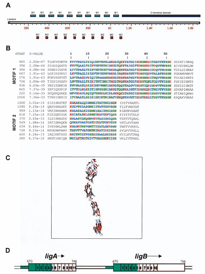
A. Schematic representation of LigB. Motif 1 (blue box) and Motif 2 (red box) comprise the 10 Big2 repetitive domains from residue 220 through 1109, whereas the C-terminus (dark blue box) is located between 1111 and 1890.
B. MEME Motif Discovery analysis of the Big2 repetitive domains of LigB. L. interrogans Lig proteins contain ~90mer repeats composed of two motifs of length 52 residues (M1) and 24 residues (M2), respectively. Residues are coloured based on their chemical properties: hydrophobic, basic, acidic and polar uncharged. The sites are listed in order of decreasing statistical significance (P-value). The P-values and start sites of the motifs are indicated. The P-value of a site is computed from the match score of the site with the position specific scoring matrix for the motif and represents the probability of a random string (generated from the background letter frequencies) having the same match score or higher.
C. Comparative modelling of L. interrogans LigB repetitive domains (residues 493–917).
D. Genetic structure of the ligAB locus. The 11 and 10 Big2 domains encoded by ligA and ligB, respectively, are numbered. Two additional N-terminal Big2 domains with poor E-values (>10−10) are not shown. The >2 kb segment of DNA sequence identity between the 5′ portions of ligA and ligB is indicated in green.