
Fig. 6.
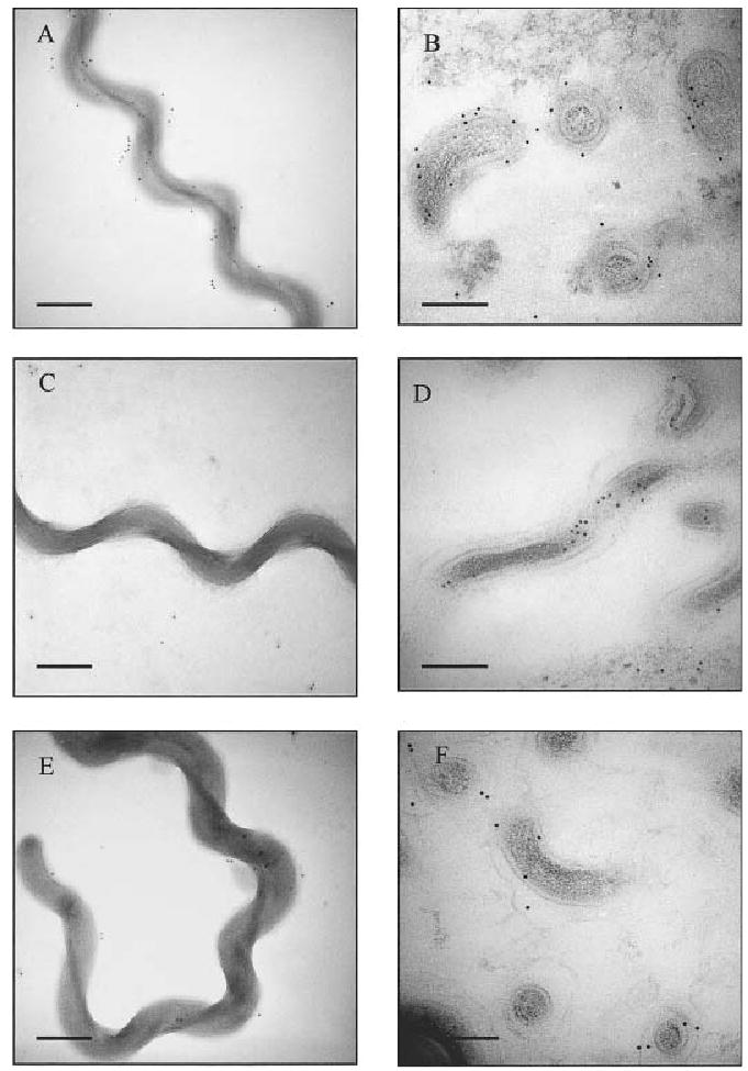
Immunoelectron microscopy of whole-cell (A, C and E) and thin-section (B, D, F) preparations of L. kirschneri strain RM52. Preparations were incubated with anti-LPS (A, B), anti-GroEL (C, D), and anti-LigB antibody (E, F), followed by anti-rabbit secondary antibody conjugated to 10 nm gold particles. Bars represent 100 nm.