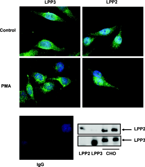
Figure 5. Subcellular distribution of endogenous LPP2 and LPP3 in CHO cells.
Serum-deprived CHO cells were stimulated without and with PMA (1 μM; 10 min). LPP3 or LPP2 expression was detected using anti-LPP3 or anti-LPP2 antibodies (green) respectively. The photograph shows re-localization of endogenous LPP3, but not LPP2, to the perinuclear compartment in response to PMA. The nuclei were stained with DAPI (4,6-diamidino-2-phenylindole) (blue). Also shown are Western blots probed with anti-LPP3 and -LPP2 antibodies to demonstrate expression of endogenous LPP3 and LPP2 in cell lysates respectively. Recombinant LPP2 and LPP3 in detergent extracts of cell membranes from stably transfected HEK-293 cells are shown as positive controls. Secondary antibody alone (Ig) was used in immunofluorescent staining experiments to show the specificity of the LPP2 and LPP3 immunoreactivity with the respective anti-LPP2 and -LPP3 antibodies.

