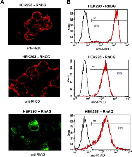
Figure 1. Cell surface expression of the recombinant RhBG, RhCG and RhAG antigens in HEK-293 transfectants.
(A) Immunofluorescence microscopy analysis. HEK-293 transfectants cultured on poly(L-lysine) coverslips were fixed and permeabilized before adding the relevant anti-Rh primary antibodies. Alexa-Fluor labelled IgG were used as secondary antibodies. (B) Flow-cytometric analysis using LA18.18 monoclonal anti-RhAG, polyclonal anti-RhBG-Cter and anti-RhCG-Cter antibodies. The black lines represent the non-transfected cells and red lines represent transfected cells. Percentage of positive transfected cells is indicated.

