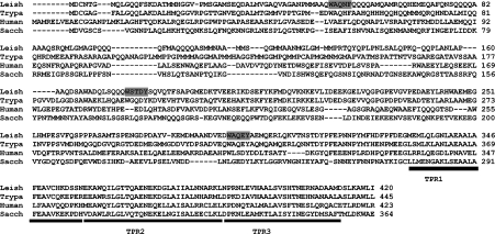
Figure 1. Multiple sequence alignment of PEX5 proteins.
A partial sequence alignment of the N-terminal region of the L. donovani (Leish), T. brucei (Trypa), human and S. cerevisiae (Sacch) PEX5 was performed using the CLUSTAL_X computer program [35]. The WXXXY/F motifs in the LdPEX5 are designated by the grey shaded boxes. The black bar delineates the first three TPRs that form part of the PTS1-binding pocket.

