
Fig. 3:
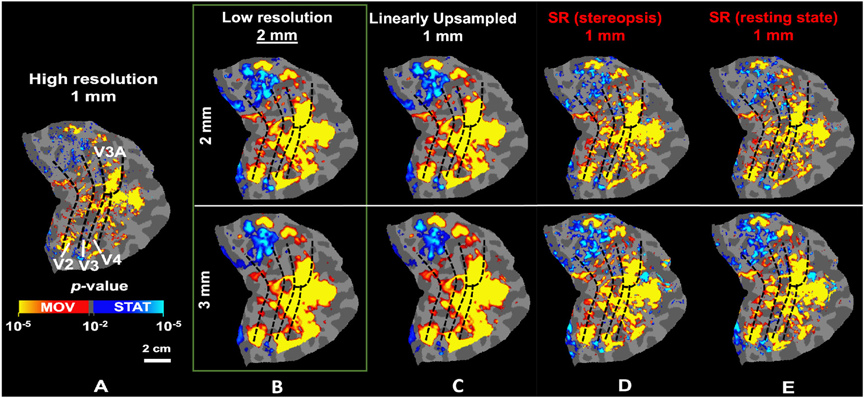
The application of the SR method improves the localization of motion-selective activity maps from low-resolution fMRI. A) Localization of motion-selective sites across V2, V3, V3A, and V4 based on the original high-resolution fMRI. B-C) Localization of the same sites based on downsampled data. Fine-scale sites are either absent or fused, causing overestimation in the size of the selective sites. D-E) Localization of motion-selective sites based on super-resolved images. The fine-scale motion-selective sites are mostly recovered in these maps. In all panels, dashed black lines indicate the borders of visual areas, defined retinotopically.