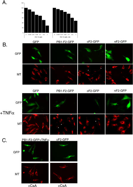
Figure 7. PB1-F2 Protein Induces Mitochondrial Permeabilization in ANT3-Dependent Manner.
(A) Recombinant PB1-F2 protein was incubated with 50 μg of purified mouse liver mitochondria in the presence or absence of 50 μM BA for 30 min. The mitochondria were further processed for assessment of membrane potential by JC-1 fluorescence at 590 nm.
(B) PB1-F2 induces loss of mitochondrial membrane potential in transfected cells. HeLa cells were transfected with GFP fusion constructs of PB1-F2, and 12 h later were treated with 50 ng/ml TNFα for 8 h, where indicated. Cells were subsequently stained with Mitotracker CMXRos Red dye. Cells with dissipated membrane potential are indicated by (*).
(C) The mitochondrial permeability transition inhibitor CsA inhibits PB1-F2-induced loss of mitochondrial membrane potential. HeLa cells in presence of CsA were transfected and treated as in (B) and stained with Mitotracker dye.

