FIG. 4.
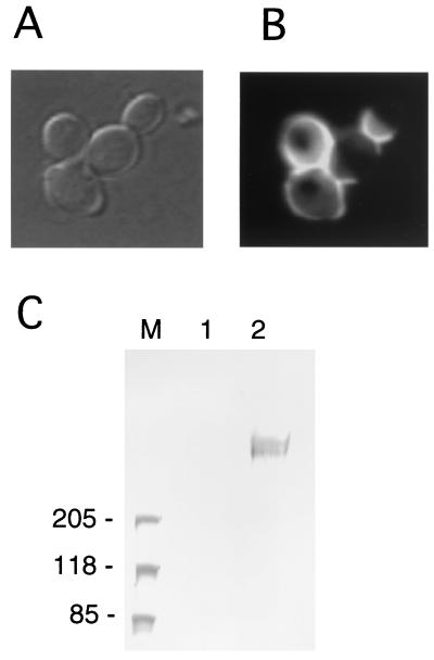
Localization of 3×HA Awa1p. (A) Representative cells of YHS235 (UT-1 harboring pRS416-XHA::AWA1) viewed under a light microscope. (B) The same field viewed with a fluorescence microscope. Fixed yeast cells were probed with the anti-HA antibody and the fluorescein isothiocyanate-labeled anti-mouse immunoglobulin G. (C) Western blotting analysis of the cell wall proteins. The cell wall fraction was prepared from yeast cells and treated with β-1,6-glucanase. Liberated proteins were analyzed by Western blotting on an SDS-2 to 15% polyacrylamide gel electrophoresis gel using the anti-HA antibody. Lane M, molecular size marker; lane 2, β-1,6-glucanase extract of YHS233 (UT-1 harboring pRS416-AWA1); lane 3, the β-1,6-glucanase extract of YHS235 (UT-1 harboring pRS416-XHA::AWA1).

