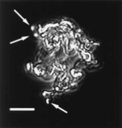
FIG. 6.
Strain PV-1 and filament formation. This pure culture was grown in liquid MWMM/ASW medium. This image is a composite of a phase-contrast image and an epifluorescence image. The preparation was stained with Syto to reveal the cells, denoted with arrows, that are attached to the Fe oxides. Note that the cells appear to grow at the termini of the filaments. Compare the morphology of these bacterially formed filaments to that of the filaments observed directly from the Loihi site (Fig. 4B).

