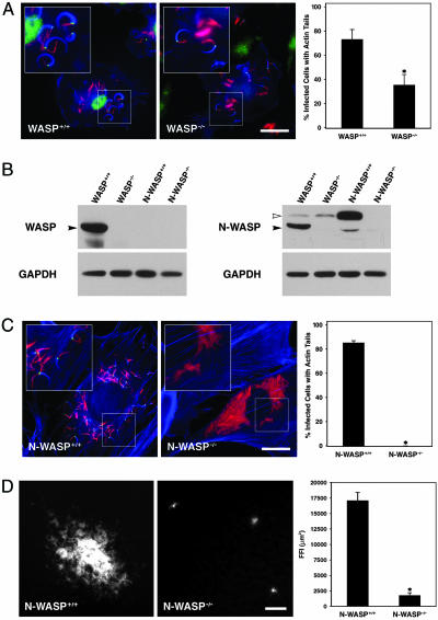
Fig. 1.
WASP or N-WASP is required for efficient M. marinum motility. (A) WASP+/+ macrophages (Left) were infected with M. marinum (red) and stained with phalloidin (blue) and with anti-WASP antibodies (green). WASP-/- macrophages (Center) were similarly infected and stained with anti-N-WASP antibodies (green). In Right, the percentage of infected cells with actin tails from three independent experiments is shown (P = 0.037). (Scale bar, 20 μm.) (B) Whole-cell lysates of WASP+/+ and WASP-/- macrophages and N-WASP+/+ and N-WASP-/- fibroblasts were resolved by SDS/PAGE and analyzed for the presence of WASP and N-WASP by Western blotting. Filled arrowheads indicate the WASP bands; open arrowheads indicate the N-WASP bands. The WASP antibody is specific for WASP, but the anti-N-WASP antibody is cross-reactive with WASP, leading to the detection of a WASP band in the WASP+/+ lane. (C) N-WASP+/+ fibroblasts (Left) and N-WASP-/- fibroblasts (Center) were infected with M. marinum (red) and stained with phalloidin (blue) and with anti-N-WASP antibodies (green). In Right, the percentage of infected cells with actin tails was quantified as in A. (Scale bar, 20 μm.) (D) N-WASP+/+ (Left) and N-WASP-/- (Center) fibroblast monolayers were infected with fluorescent M. marinum, and foci of infection were visualized and measured 8 days later (P = 0.002). (Scale bar, 50 μm.) Asterisks in A-D indicate statistically significant differences with P < 0.05.

