Figure 1.

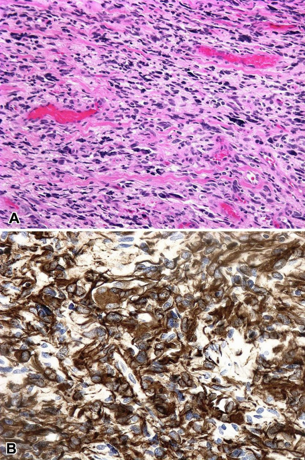
A. Biopsy of an intradural extramedullary lesion at T12/cauda equina, showing highly pleomorphic cells with a hint of fibrillary cytoplasm growing in an infiltrating fashion (H&E, ×100). B. Diffuse cytoplasmic staining of tumor cells with antibody for GFAP (GFAP, ×200).
