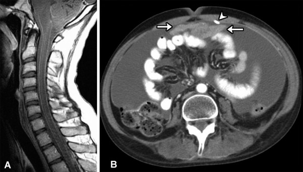
Figure 2.

A. Postcontrast sagittal T1-weighted MR image of the cervical spine shows diffuse leptomeningeal enhancement along the surface of the spinal cord. B. Axial contrast-enhanced CT image through the abdomen demonstrates extensive ascites and an enhancing midline mass (arrows) adjacent to the shunt catheter (arrowhead).
