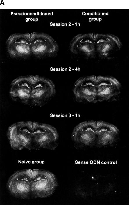
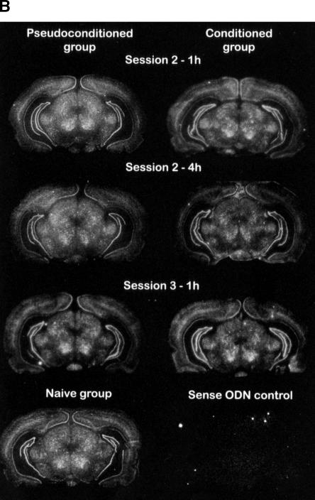
Figure 2.
Representative distribution of Kv1.1 mRNA in the rat hippocampus of three experimental groups, i.e., naive, pseudoconditioned, and conditioned groups. (A) Dorsal hippocampus; (B) ventral hippocampus. The rats were sacrificed 1 or 4 h following session 2 or 1 h following session 3. Kv1.1 mRNA signals were revealed by autoradiographic in situ hybridization using a specific antisense probe labeled with 35S. Brain regions were defined according to Paxinos and Watson (1986). White pixels represent a high mRNA-labeling level, while black pixels, no labeling.


