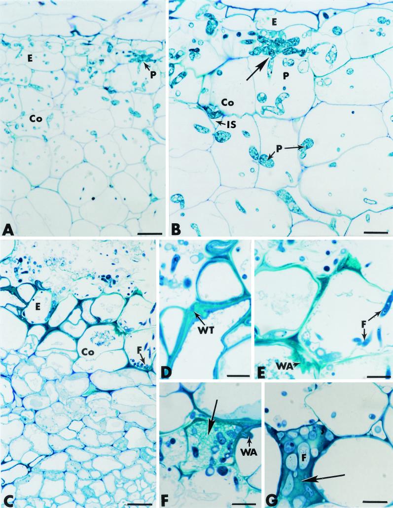
FIG. 5.
Cross sections of pea roots stained with toluidine blue. (A and B) Roots inoculated with P. ultimum (controls). Hyphae of P. ultimum (P) propagate through much of the root tissues, including the epidermis (E) and the cortex (Co) (panel A). Fungal growth occurs in intercellular spaces (IS), as well as intracellularly (panel B). Direct host cell wall penetration is visible (panel B, arrow). Bars: A, 25 μm; B, 10 μm. (C through G) Roots inoculated with F. oxysporum strain Fo47. The fungus (F) develops actively at the root surface and penetrates the epidermis (E) and the outer cortex (Co) (panel C). Colonization of the outermost root tissues correlates with marked thickening of the host cell walls (panel D), formation of wall appositions (panel E), deposition of a fibrillar matrix in which fungal cells (F) are trapped (panel F, arrow), and occlusion of intercellular spaces with amorphous material in which fungal cells are also captured (panel G, arrow). Bars: C, 25 μm; D through G, 10 μm.

