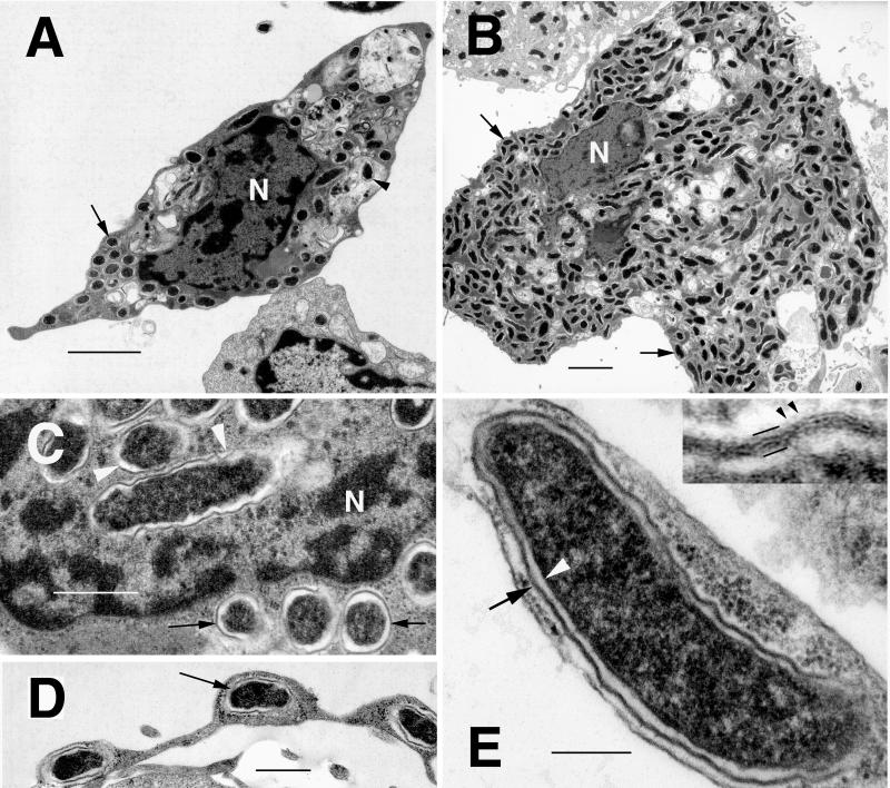
FIG. 1.
Transmission electron photomicrographs of R. monacensis IrR/MunichT within cultured tick and mammalian cells. (A) Infected I. scapularis (ISE6) cell with rickettsiae in the cytoplasm (arrow), as well as being digested in vacuoles (arrowhead). Bar = 2 μm. N, host cell nucleus. (B) Vero cell filled with cytoplasmic rickettsiae (arrows). N, host cell nucleus. Bar = 2 μm. (C) R. monacensis within the nucleus (N, arrowheads) and cytoplasm (arrow) of an ISE6 cell. Bar = 0.5 μm. (D) Rickettsia (arrow) within a pseudopodial extension of an ISE6 cell. Bar = 0.5 μm. (E) High magnification of R. monacensis showing the typical rickettsial morphology of the organism. Bar = 0.2 μm. Shown are the inner periplasmic membrane (white arrowhead), the electron-lucent periplasmic space, and the cell wall (arrow). The insert shows an enlargement of the outer membrane. Bars delineate the trilaminar cell wall; small arrowheads indicate the faint microcapsular layer.

