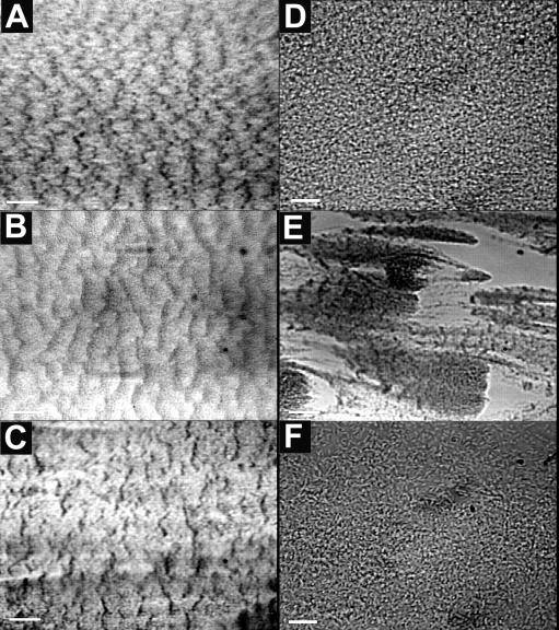
FIG. 4.
Ripple structures formed in PAO1 and JP1 biofilms. Images of PAO1 biofilm ripple structures in the biofilms growing in the turbulent flow cell (A) and the laminar flow cell (B), taken at days 4 and 5, respectively, are shown. The ripples were aligned perpendicularly to the flow direction (right to left). Scale bar, 200 μm. (C) JP1 biofilm ripple structures in the turbulent flow cell taken on day 6. Scale bar, 200 μm. The ripple structures were much less evident under higher magnification (the images in panels D and F are of the same fields as those in panels A and C, respectively). Scale bar, 20 μm. (E) Patchy PAO1 biofilm structures in the turbulent flow cell, run 3, day 5. Scale bar, 100 μm.

