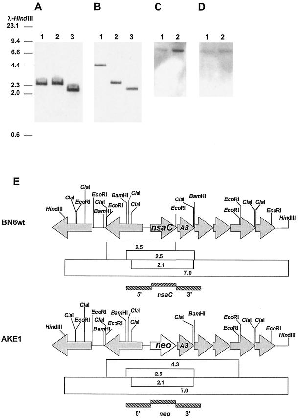
FIG. 1.
Southern blot analysis of chromosomal DNA of strains AKE1 and BN6wt. DNA was digested, electrophoresed on a 0.7% agarose gel, transferred to a nylon membrane, and hybridized. HindIII-digested λ DNA was used as a marker. (A) Southern hybridization of chromosomal DNA of BN6wt digested with different restriction enzymes using the nsaC gene fragment as the probe. Lane 1, EcoRI; lane 2, BamHI; lane 3, ClaI. (B) Southern hybridization of chromosomal DNA of strain AKE1 digested with different restriction enzymes using the neo gene fragment as the probe. Lane 1, EcoRI; lane 2, BamHI; lane 3, ClaI. (C) Hybridization of HindIII-digested chromosomal DNA using the 1-kb 5′ flanking sequence of nsaC as the probe. Lane 1, strain AKE1; lane 2, strain BN6wt. (D) Hybridization of HindIII-digested chromosomal DNA using the 1-kb 3′ flanking sequence of nsaC as the probe. Lane 1, strain AKE1; lane 2, strain BN6wt. (E) Restriction map of the genomic DNA of these strains. The position of the nsaA3 gene encoding the ferredoxin subunit of the NSDO which was identified adjacent to nsaC (26) is shown. The sizes (in kilobases) of restriction fragments detected by Southern hybridization are indicated in open boxes. DNA fragments representing the probes used for hybridization are indicated by shaded boxes.

