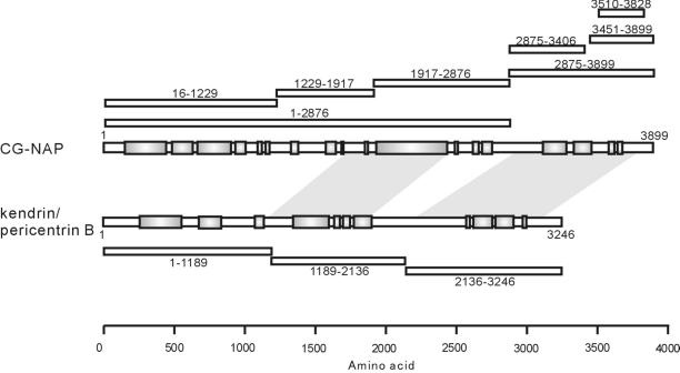
Figure 1.
Schematic representation of CG-NAP, kendrin, and their deletion mutants. Schematic structure of CG-NAP and kendrin are shown with predicted coiled-coil regions in shaded boxes. Positions of the deletion mutants of CG-NAP and kendrin are shown with amino acid residues on the upper and lower sides, respectively. Shaded areas between CG-NAP and kendrin represent the regions sharing homology found by BLAST search. Total amino acid residue of kendrin used in this study was 3246, as described in MATERIALS AND METHODS, which is shorter than that (3321) deposited to GenBank with accession number U52962.

