Figure 4.
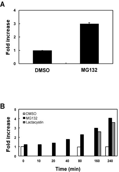
Proteasomal inhibitors increase cell surface mLRP4. (A) CHO-mLRP4 cells were treated with DMSO or DMSO containing MG132 (20 μM) for 2 h. Cells were then dissociated and immunostained with anti-HA antibody. Cell surface mLRP4 was detected with goat anti-mouse Ig-FITC and FACS analysis. Background fluorescence intensity was assessed in the absence of primary antibody and subtracted from each assay. The mean values from representative triplicate determinations were averaged with the SE value given as error bars. (B) Kinetic analyses of cell surface mLRP4 after treatment with proteasomal inhibitors. CHO-mLRP4 cells were treated with MG132 (20 μM) for various periods of time as indicated. Treatment with the alternative proteasome inhibitor lactacystin (20 μM) or vehicle DMSO alone were also performed at selected time points. Cell surface mLRP4 was determined via FACS analysis as indicated above. Shown in the figure are results of a representative experiment. Note the gradual increase of cell surface mLRP4 upon treatment with proteasomal inhibitors, but not DMSO.

