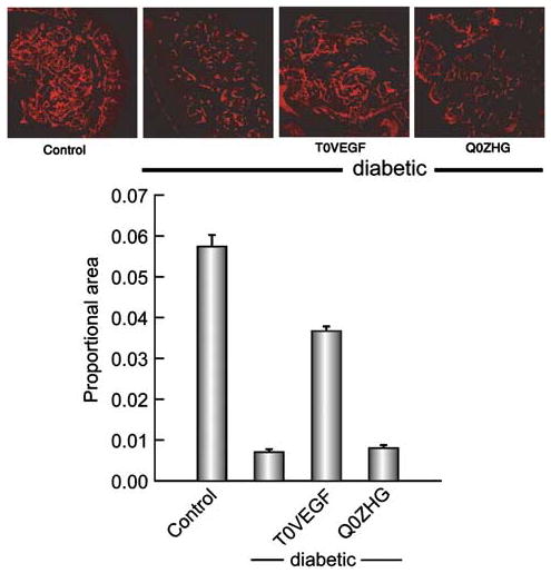
Figure 6.

Nerve fibers in the footpad visualized by PGP 9.5 immunoreactivity. The nerve area, quantitated as proportional area occupied by the nerve in the footpad, was substantially reduced in diabetic animals but preserved in diabetic animals inoculated with T0VEGF. Mean±s.e.m.; * P<0.005 compared to diabetic or diabetic animals inoculated with Q0ZHG; n = 5 animals per group.
