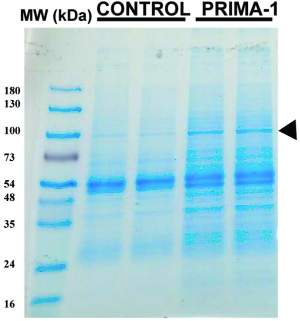
Figure 1.

Coomassie blue-stained gel of proteins co-immunoprecipitated with DO-1 primary antibody from MDA-MB-231 cells. Cleared cell lysates were immunoprecipitated with DO-1 primary antibody directed against p53, washed, and resolved by SDS-PAGE (4 to 20% polyacrylamide). Two independent co-immunoprecipitated samples from untreated control (-) and cells treated for 4 hours with 100 μM PRIMA-1 (+) were loaded. The gels were stained with Coomassie blue. Molecular masses of protein size markers are indicated (MW). The arrowhead indicates the band of stained proteins excised for enzymatic digestion by trypsin and subsequent mass fingerprinting with matrix-assisted laser desorption ionization-time-of-flight mass spectrometry.
