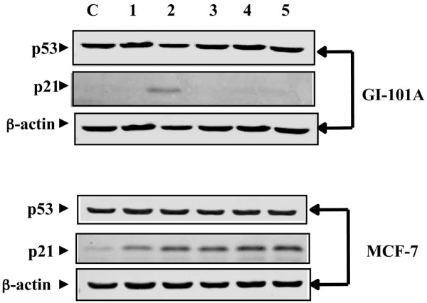
Figure 2.

Inhibition of PRIMA-1 mediated transcriptional reactivation function of p53 with pifithrin-α (PFTα). MCF-7 (p53+/+) and GI-101A (mut p53) cells were treated with 100 μM PRIMA-1 for 2, 4 and 8 hours (lanes 1, 2 and 3, respectively). Cells were treated with 20 μM PFTα for 6 hours (lane 4) or with 20 μM PFTα for 2 hours followed by PRIMA-1 for 4 hours (lane 5). 20 μg of protein samples of cell lysates were separated by SDS-PAGE (4 to 20% polyacrylamide) and subjected to Western blot analysis with p53 and p21 primary antibodies. The reactive bands were revealed and detected with the Odyssey™ Infrared Imaging System. β-Actin was used as a loading control for protein samples.
