
Figure 1.
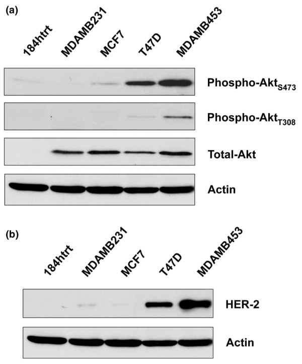
Examination of P-Akt and HER-2 levels in a panel of breast cancer cell lines. (a) Proteins were isolated from cells growing in log phase and the levels of phosphorylated Akt (P-Akt) were then assessed using antibodies to serine 473 and threonine 308. MDA-MB-453 cells expressed the highest levels of activated Akt relative to the other lines. There were no differences in the levels of total Akt with the exception of the preneoplastic cell line 184htrt. Actin was detected as a loading control. (b) Human epidermal growth factor receptor (HER)-2 protein expression was evaluated in a panel of breast cell lines. The MDA-MB-453 and T47D cells expressed HER-2 whereas the other cell lines did not.