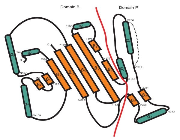
Figure 4.
Topology diagram showing the arrangement of secondary-structure elements in the FBG domains of TL 5A. Domains named in analogy to human fibrinogen γ chain fragment. α-helix is represented in green and β-sheet is represented in brown. Domain B and domain P are separated by a red line. Starting position of amino acid in each secondary structure is shown in the figure with single letter. The disulfide bridge (Cys-206-Cys-219) in the domain P is represented by a dot line.

