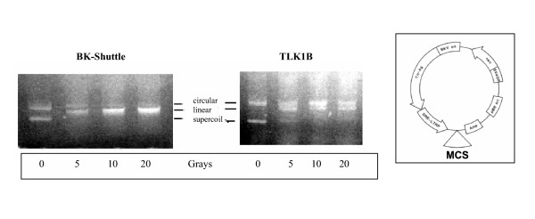
Figure 4.
Analysis of episomes. 2 × 10⁷ cells transformed with BK-Shuttle or TLK1B were irradiated with the indicated dose of γ-radiation. The cells were returned to the incubator and the plasmids were isolated 1 hr later by the Hirt's protocol and separated on a 1% agarose/TAE gel. The mobility of the forms (circular, linear, and supercoiled) is indicated. The structure of the BK-Shuttle episomal vector is shown on the right. The bands were quantified with ImageQuant vs. 5 (Molecular Dynamics).

