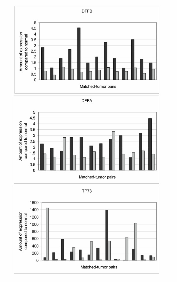
Figure 2.
QRTPCR results in oligodendroglioma subsets. Black bar, 1p-intact samples; light gray bars, 1p-loss samples. A) Expression of DFFB was lower in all 1p-loss samples as compared to their matched 1p-intact samples. Ten of the thirteen 1p-loss tumors had lower expression of DFFB compared to normal brain, with three tumors demonstrating 1–2× the amount of normal brain DFFB expression. In contrast, all 1p-intact tumors had DFFB expression greater than or equal to that seen in normal brain. B) Expression of DFFA was lower in most 1p-loss samples as compared to their matched 1p-intact samples. Only three 1p-loss tumors had higher DFFA expression. Differential expression was detected to a degree; most 1p-loss tumors (10 of 12) demonstrated 1–2× the amount of normal brain DFFA expression, whereas most 1p-intact tumors (9of 12) demonstrated ≥ 2× the amount of normal brain DFFA expression. C). Differential p73 expression was not detected. In five of the pairs, 1p-loss tumors had higher expression, whereas in seven other pairs, 1p-intact tumors had higher expression.

