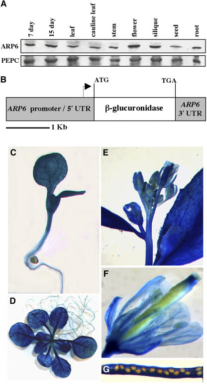
Figure 3.
Organ- and Tissue-Level Expression Patterns of ARP6.
(A) Immunoblot of protein extracts from whole plants (7 day, 7-d-old seedling; 15 day, 15-d-old plant) or various plant organs probed for the 47-kD ARP6 with the monoclonal antibody mAbARP6a (top panel) and for the 115-kD PEP-carboxylase with anti-PEP carboxylase (PEPC, bottom panel) polyclonal antibody as a loading control. Each lane was loaded with ∼25 μg of total protein, and the two panels shown were from different segments of the same blot. The flower protein sample was prepared from the entire inflorescence tip and thus contains flowers of all stages.
(B) Diagram of the GUS coding sequence under control of the ARP6 regulatory sequences. The ARP6 promoter/5′ untranslated region (UTR) is defined as the 2-kb region upstream of the ARP6 start codon, and the ARP6 3′ UTR consists of the 400-bp region downstream of the ARP6 stop codon.
(C) to (G) Transgenic plants carrying the reporter transgene shown in (B). Seven-day-old seedling (C); 20-d-old plant (D); inflorescence tip showing flowers, stems, and cauline leaves (E); a single flower (F); mature green silique opened to expose seeds (G).

