Figure 2.
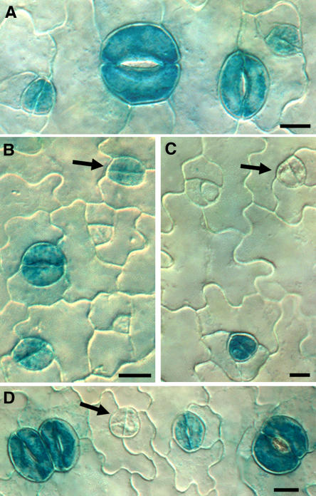
Fewer GMC Daughter Cells Show Guard Cell–Specific ProKAT1:GUS Staining in flp-1.
(A) Wild type. GUS staining present in developing and mature stomata.
(B) Wild type. Arrow indicates staining in daughter cells produced by GMC division. Staining is absent from stomatal precursors (GMCs and meristemoids) in both the wild type and flp-1.
(C) and (D) flp-1. Arrows indicate daughter cells without staining that are likely to function as GMCs and divide one more time.
Bars in (A) and (B) = 7.5 μm; bars in (C) and (D) = 10 μm.

