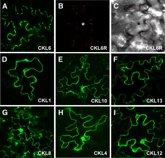
Figure 7.
Arabidopsis CKL6 Localizes to Discrete Regions at the Cell Periphery of Tobacco and Arabidopsis Epidermal Cells.
Tobacco leaf tissue transfected using Agrobacterium tumefaciens transformed with GFP-tagged CKL isoforms ([A] and [D] to [I]) was analyzed 48 h after agroinfiltration. Arabidopsis leaf tissue biolistically bombarded with plasmid DNA expressing RFP-tagged CKL6 ([B] and [C]) was analyzed 24 h after biolistic DNA delivery. Confocal microscopic images represent paradermal regions of epidermal cells expressing these fluorescently tagged constructs.
(A) CKL6-GFP develops a punctate fluorescence pattern reminiscent of plasmodesmal pit field distribution within epidermal cell walls.
(B) CLK6-RFP signal in an Arabidopsis epidermal cell shows punctate fluorescence signals similar to those observed in tobacco cells.
(C) Same image shown in (B) but combined with the transmitted light image to show the contour of the bombarded cell (indicated by an asterisk).
(D) to (F) CLK1-, CLK10-, and CLK13-GFP exhibit punctate fluorescence signals.
(G) CLK8-GFP signal appears to label cytoplasm and nucleus and to form punctate particles.
(H) CKL4-GFP signal accumulates in the nucleus and cytoplasm; note the absence of a punctate pattern over the cell wall.
(I) CKL12-GFP signal was strongest in the cytoplasm but was also present in the nucleus; note the absence of a punctate pattern over the cell wall.

