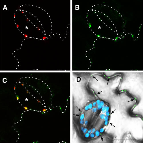
Figure 8.
Colocalization of PAPK1 and TMV MP in Symplasmically Isolated Guard Cells.
PAPK1-RFP was transiently expressed, by particle bombardments, in transgenic tobacco leaves expressing TMV MP-GFP.
(A) Fluorescent signals, produced by PAPK1-RFP in a target guard cell (asterisk), detected in the red channel of the confocal microscope. Contours of the epidermal cells, drawn with white dashed lines, allow identification of the target cell in the context of the epidermal layer.
(B) Fluorescent signals, produced from TMV MP-GFP, were observed in the periphery of all epidermal cells, detected in the green channel on the confocal microscope. Note that the strong accumulation of TMV MP-GFP signal is only detected in one guard cell that ectopically expressed PAPK1-RFP, which is in contrast with the neighboring guard cell in which TMV MP-GFP was not detectable.
(C) Combined image of (A) and (B) showing overlapping signals between the red and green channels. Note the presence of strong yellow signals confined to the bombarded guard cell.
(D) Control image showing guard cells and surrounding epidermal cells in a TMV MP-GFP transgenic tobacco leaf. Picture represents a combined image of signals collected from three different channels. Punctate green signal, representing TMV MP-GFP, accumulates in plasmodesmata (arrows). Note that hardly any signals are detected within guard cells. Blue signal (false color): chloroplast autofluorescence restricted to the guard cell pair. Gray image: epidermal cells collected in the transmitted-light channel. Bar = 20 μm for (A) to (D).

