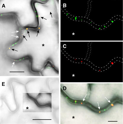
Figure 9.
PAPK1 and TMV MP Colocalize within the Cross-Walls of Neighboring Epidermal Cells.
Leaves of a TMV MP-GFP–expressing transgenic tobacco line were bombarded with PAPK1-RFP plasmid and observed by confocal microscopy 24 h after bombardment.
(A) Overlapping TMV MP-GFP and PAPK1-RFP signals (yellowish-orange; white arrows) detected within epidermal cross-walls; additional overlapping signals are also detected near the cell periphery (black arrows). Image represents the combination of green, red, and transmitted-light channels. Asterisk indicates the bombarded epidermal cell that is connected to its neighbors by plasmodesmata.
(B) to (D) High-resolution images show overlapping signals at the cross-wall of neighboring epidermal cells. Green channel, TMV MP-GFP (B); red channel, PAPK1-RFP (C); combined image demonstrates the overlapping nature of signals (D). Note the confinement of strong yellow-orange signals over the target cell wall (asterisk). Dashed lines in (B) and (C) illustrate the cell contour, whereas dotted lines indicate the position of a cross-wall slightly out of the focal plane.
(E) Epidermal cells imaged at lower magnification by transmitted light; boxed region represents the area shown in (B) to (D).
Bars = 10 μm in (A), 5 μm in (D), and 20 μm in (E). Bar in (D) is common to (B) and (C).

