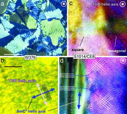
Fig. 4.
Polarized light micrographs of the TGBC phase of GBTGB materials in transmission. The TGB helix axis is indicated in white and the SmC helix in blue. (a) Grandjean texture of a 2-μm-thick cell of W376 in the GBTGBC phase, showing areas of Δ= 60° and 90° periodicity. (b) Planar-aligned W376 with the TGB and SmC helices in the plane of the cell plates, showing orthogonal quasiperiodic arrays of lines from both helices. (c) Four-micrometer cell of S1014/CE8 clearly showing the SmC helix. The dark domain to the left contains a single smectic block (bookshelf alignment) that splits into two blocks on the right, rotated ≈60° from one another. This Δ is preferred, but only weakly, as d shows, with 60° domains merging into 90° domains over ≈100 μm. (Scale bars: 20 μm, a and b; 10 μm, c and d.)

