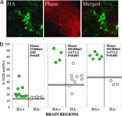
Fig. 5.
Expression of Mito-ApaLI-HA in brain leads to a shift in mtDNA heteroplasmy. The putamen of anesthetized 2- to 4-mo-old mice was stereotactically injected with 2 μl of virus suspensions (Ad5-Mito-ApaLI-HA or AAV1,2-Mito-ApaLI-HA). After 1 (Ad5) or 2 weeks (AAV1,2), the animals were killed and the brains snap-frozen in liquid nitrogen. Twenty-micrometer sections were screened for GFP expression (not shown) and adjacent sections stained for HA expression (a). Regions of positive and negative staining were microdissected by laser capture microscopy and subjected to mtDNA haplotype analysis as described in the legend to Fig. 4b. Horizontal gray bars represent the percent NZB mtDNA in the mouse tail.

