Fig. 2.
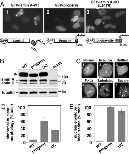
Expression of GFP-progerin and GFP-lamin A-UC in HeLa cells results in abnormal nuclear morphology and a localization pattern distinct from GFP-lamin A-WT. (A) HeLa cells were transiently transfected with the indicated constructs (containing the L367P mutation) as described in Materials and Methods; representative fields of cells are shown. Schematics of the expected processing status of the GFP-tagged proteins are shown. (B) Expression of the GFP-tagged proteins (arrow) and endogenous lamin A and C was assessed by Western blotting with anti-lamin A/C antibodies (Upper), with tubulin as a loading control (Lower). (C) The different types of normal (image 1) or aberrant (images 2-6) nuclei observed in cells expressing GFP-progerin, ranging from mild to severe, are indicated. (D and E) Nuclei from HeLa cells transfected with the GFP-tagged constructs were scored for abnormal nuclear morphology (D) and nuclear envelope localization (E) of the GFP-tagged proteins as described in Materials and Methods. Graphs represent the average of two experiments with the range indicated by the bar.

