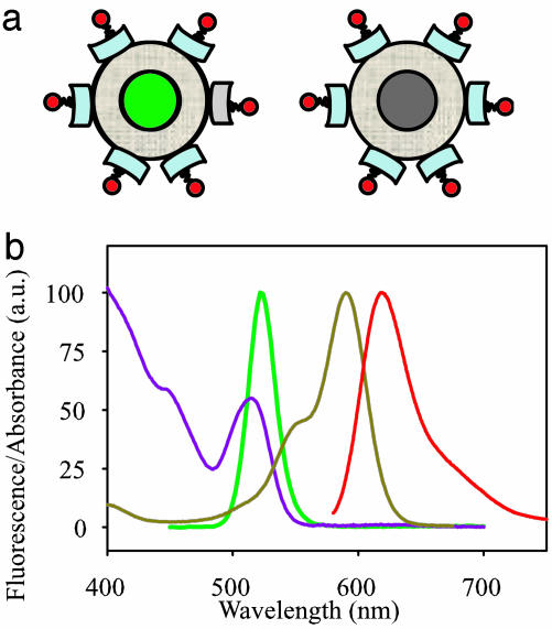
Fig. 1.
Schematic structure and spectra of qdot particles used in the experiments. (a) Qdot-SAv (central sphere of CdSe/ZnS core/shell, tan outer layer of amphiphilic coating for water solubility, and light blue block of SAv) labeled with biocytin-Alexa Fluor 594 (red). (Left) Bright (green) qdot. (Right) Dark (gray) qdot. The cartoon does not indicate the actual dye-loading ratio. (b) Normalized fluorescence absorption and emission spectra of Alexa Fluor 594 (dark yellow and red, respectively) and of Qdot525-SAv (purple and green, respectively).

