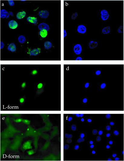
Figure 6.
Nuclear localization of the L- and D-forms of the F3 peptide. Nuclear localization in HL-60 cells of F3 (a) or ARALPSQRSR control peptide (b). (c–f) Uptake by MDA-MB-435 cells of F3 synthesized either from L or D amino acids. The cells were treated as in Fig. 5A and stained with 4′,6-diamidino-2-phenylindole before examination under a confocal (a and b) or inverted fluorescent microscope (c–f). (c and e) Peptide staining (green); (d and f) nuclear staining (blue). (Magnifications: a and b, ×400; c–f, ×200.)

