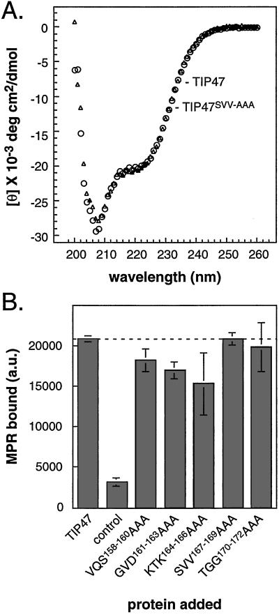
Figure 2.
(A) CD analysis of TIP47 (circles) and TIP47SVV-AAA (triangles). Units for mean residue ellipticity [θ] are degrees·cm2⋅dmol−1. (B) TIP47 and TIP47 mutants are fully active in binding MPR cytoplasmic domains. His-tagged TIP47 (200 nM) was incubated with GST–CI-MPRΔ12. Complexes were recovered on nickel beads and analyzed for the presence of GST–CI-MPR by immunoblot. Backgrounds were determined as in Fig. 1B and were subtracted. Shown are means and standard deviations of duplicate samples. Results are representative of three independent experiments. The dashed line is included as in Fig. 1.

