Figure 3.
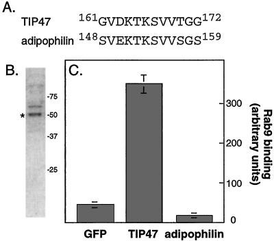
(A) Sequence comparison of TIP47 and adipophilin. (B) Coomassie blue stained-SDS/PAGE analysis of purified adipophilin (*); adipophilin is the lower band in the doublet labeled, as determined by immunoblot. (C) Adipophilin does not bind Rab9. Binding was carried out as described in Methods; error bars represent the SE for duplicate measurements.

