Figure 1.
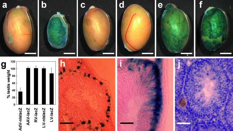
Transduction performance of viral vectors in testicular cells. We injected viral vectors into the seminiferous tubule of B6D2F1 wild-type testis and analyzed at 2 mo after injection. Whole testis was fixed and stained with X-Gal to observe lacZ reporter gene expression. (a) Wild-type control. (b) Adenoviral vector (AdV-CMV-nlslacZ). (c) Adeno-associated viral vector (AAV-CMV-lacZ). (d) Retroviral vector (RV-CMV-lacZ). (e) Lentiviral vector (LV-CMV-nlslacZ). (f) Lentiviral vector (LV-CMV-lacZ). (g) The values are percent testes weight (injected right testis/uninjected left testis, mean ± SD) at 2 mo after injection. AdV-CMV-nlslacZ; 36.1 ± 10.2 (n = 6), AAV-CMV-lacZ; 102.0 ± 3.5 (n = 3), RV-CMV-lacZ; 101.6 ± 6.4 (n = 4), LV-CMV-nlslacZ; 100.9 ± 4.6 (n = 7), LV-CMV-lacZ; 86.8 ± 14.8 (n = 4). (h and i) X-Gal-stained frozen testis cross sections. Nuclear localization and cytoplasmic diffusion of β-galactosidase in Sertoli cells were observed with LV-CMV-nlslacZ (h) and LV-CMV-lacZ (i), respectively. (j) Morphologically normal spermatogenesis was observed in the testis injected with LV-CMV-nlslacZ. [Bar = 2 mm (a–f) and 50 μm (h–j).]

