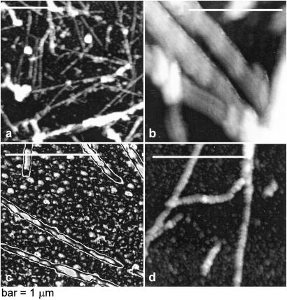
Figure 5.
Atomic force microscopy images of the deoxy-HbS polymers formed at 35°C in a 22-g/dl solution of deoxy-HbS. (a) Low-resolution image showing a variety of polymeric structures. (b–d) High resolution images showing thicker fibers consist of thinner polymers of 22 nm diameter (b). (c) The chosen color scheme highlights the pitch of the thin polymer fibers of ∼150 nm. (d) Branched and twisted polymer fibers. Under these conditions, observations of such structures were relatively rare.

