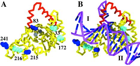
Figure 2.
(A) The structure (PDB ID code 1EXN) determined for T5 5′ nuclease showing the helical arch (red backbone), divalent metal ions (gray spheres), and space-filling representations of selected lysine (blue) and arginine (cyan) residues. (B) Original DNA-binding model proposed by Ceska et al. (8). The duplex parts (I and II) of the substrate lie across a slightly concave and positively charged region of the protein. In this model residues K241, K215, and R216 contact duplex I, and R172 contacts duplex II.

