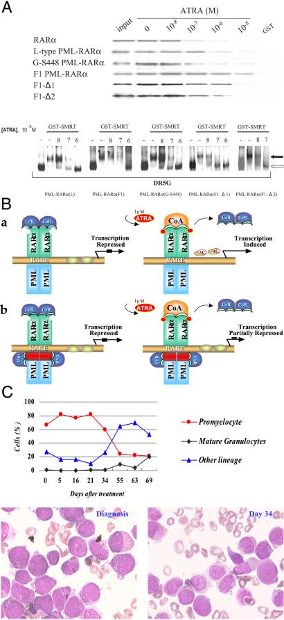
Figure 3.
(A Upper) Ligand-dependent dissociation of SMRT from wild-type RARα and different PML-RARα proteins. (A Lower) Gel-shift analysis of the interaction between GST-SMRT and the L-type, F1, F1-Δ1, F1-Δ2, and G-S448 PML-RARα on DR5G and the effect of ATRA. The filled and open arrows indicate the bands of PML-RARα/SMRT complex and PML-RARα homodimer, respectively. (B) A molecular model of APL pathogenesis and differentiation therapy. Under physiologic concentrations of ATRA, the CoR complex still binds with typical (a) or V-type (b) PML-RARα, thereby repressing target gene transcription. When APL cells are treated with 10−6 M of ATRA, this repression can be relieved in typical PML-RARα (a) but not in F1 V-type, because of a second CoR binding site in the spacer (red). (C) Clinical response of the F1 case to ATRA. (Upper) The changes of cellular components in bone marrow (BM). (Lower Left and Right) BM picture at diagnosis and on day 34 of ATRA therapy, respectively.

