Figure 5.
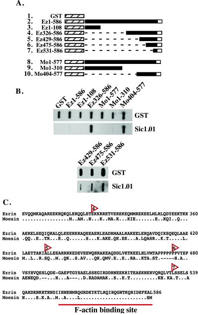
Sic binds to the carboxyl terminus of ezrin and moesin. (A) Schematic of ezrin and moesin GST fusion proteins used for immunoblot analysis. Ez1–586, full-length GST-ezrin fusion construct; Mo1–577, full length GST-moesin fusion construct; hatched box, GST; black box, ezrin or moesin; white box, F-actin binding site in the carboxyl terminus of ezrin or moesin; dashed lines, deleted amino acids. (B) GST or the indicated GST fusion proteins were purified in the absence of detergent and 1 μg of each protein vacuum-blotted to nitrocellulose. Blots were incubated with 20 μg of Sic1.01, and bound Sic was detected with specific antibody. Transferred GST fusion proteins were detected on a duplicate blot with anti-GST antiserum. (C) Alignment of the carboxyl termini of ezrin and moesin. The alignment starts at residue 301 of ezrin (National Center for Biotechnology Information accession no. 4507893). Periods indicate identical amino acids, and dashes indicate sequence gaps. Red arrowheads indicate the starting residue of the carboxyl-terminal ezrin GST fusion proteins that bind Sic. Numbers within the arrowheads correspond to row numbers in A.

