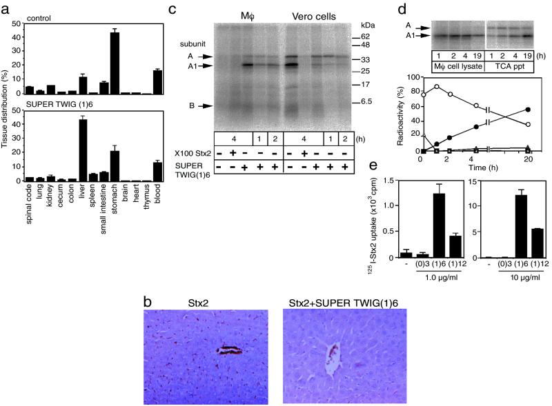
Figure 4.
SUPER TWIG (1)6-dependent degradation of Stx2 in macrophages. (a) Tissue distribution of 125I-Stx2. The data are presented as the percentage of the total radioactivity present in all of the tissues examined (means ± SE, n = 3). Total radioactivities for control and SUPER TWIG (1)6-coinjected mice were 8,900 ± 400 and 12,200 ± 2,200 cpm (means ± SE, n = 3), respectively. (b) Immunostaining of Stx2 in the liver. Stx2 present in the sections was detected by using specific antibody against Stx2. (c) Metabolism of 125I-Stx2 in cells. Mouse peritoneal macrophages (Mφ) or Vero cells were incubated with 125I-Stx2 (1 μg/ml) in the absence or presence of SUPER TWIG (1)6 (10 μg/ml) or a 100-fold excess of nonradioactive Stx2 for the indicated periods at 37°C. These lysates were separated by electrophoresis on SDS/15% polyacrylamide gel and bands visualized by autoradiography. (d) Degradation of Stx2 in macrophages. Open triangle and open circle indicate A subunit and A1 fragment, respectively, present in the macrophage cell lysate. Filled square and filled triangle indicate A subunit and A1 fragment, respectively, present in the TCA precipitate (ppt) of the culture medium. Because of the low protein content in the TCA ppt fraction, an 8 times larger aliquot of it was used for SDS/PAGE than the amount of the macrophage cell lysate used. Filled circles indicate TCA-soluble supernatants. The data are presented as the percentage of the total radioactivity present in the cell lysate and the culture medium. (e) Effect of SUPER TWIGs on the uptake of 125I-Stx2 by macrophages. Mouse peritoneal macrophages were incubated with 125I-Stx2 (1 μg/ml) in the absence or presence of SUPER TWIG (0)3, (1)6, or (1)12 (1 or 10 μg/ml) for 30 min at 37°C. The data are presented as the total radioactivity (cpm) present in the cell lysate (means ± SE, n = 3).

