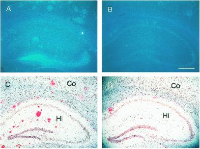
Figure 2.
Reduced deposition of amyloid plaques in hAPP+:ZnT3−/− mouse brains. (A–D) Coronal sections of 24-month-old female hAPP+ mouse brains with ZnT3+/+ (A and C) or ZnT3−/− (B and D) genotype, stained with TFL-Zn (A and B) or Congo red (C and D). Compared with ZnT3+/+ mice that had numerous TFL-Zn- and Congo red-stained plaques in cerebral cortex (Co) and hippocampus (Hi), ZnT3−/− mice had markedly reduced number of plaques. (Bar = 100 μm.)

