Figure 3.
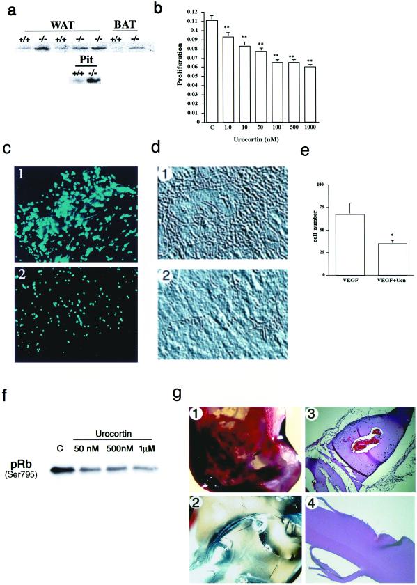
Mechanism of CRFR2 inhibition of vascularization. (a) Western blot showing increased VEGF expression in white (WAT) and brown (BAT) adipose tissue and pituitary gland (Pit) from CRFR2-mutant mice. Figure is a representative of three repeated experiments. (b) Inhibition of SMC proliferation and capillary tube formation with Ucn treatment. SMCs treated for 48 h with Ucn demonstrated a dose-responsive and significant inhibition of SMC proliferation. C, control. Data are expressed as mean ± SEM, n = 20; ∗∗, P < 0.001. (c–e) Ucn treatment also inhibited the formation of capillary-like tubes in three-dimensional collagen gels containing rat aortic endothelial cells. Formation of elongated bifurcating tubules that pervaded the gel matrix (c1 and d1) was inhibited with Ucn treatment, as a decrease in the number and size of tubules formed was seen (c2 and d2). (c) 4′,6-Diamidino-2-phenylindole (DAPI) immunofluorescence of ECs in sections from collagen gels, treated with VEGF (1) or VEGF plus Ucn (2). (×10.) (d) Phase-contrast images from collagen gels, treated with VEGF (1) or VEGF plus Ucn (2). (×10.) (e) Quantification of cell numbers in gel sections after treatment. Data are expressed as mean ± SEM, n = 3; ∗, P < 0.05. (f) Western blot showing a dose-responsive decrease in phosphorylation of Rb in SMCs after Ucn treatment. Cells were treated for 24 h before harvest. C, control. (g) Matrigel plugs demonstrating immense vascularization in the bFGF treatment (1) and no vascularization in the Ucn and bFGF treatment (2). (3 and 4) Hematoxylin and eosin-stained sections from 1 and 2, respectively showing capillary formation and EC migration (blue-stained nuclei) in the bFGF treatment (3) and inhibition of vessel formation and migration in the Ucn and bFGF treatment (4). (×10.)

