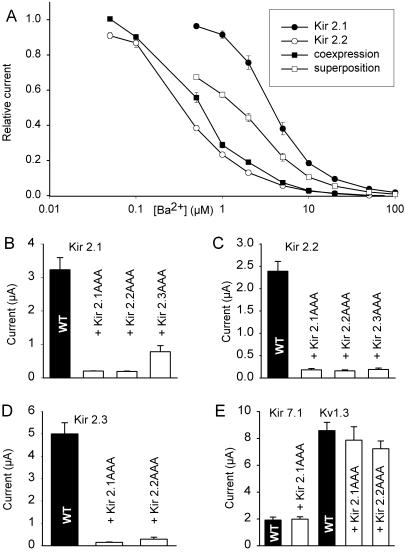
Figure 2.
Coexpression of Kir2 channels in Xenopus oocytes. (A) Concentration dependence of Ba2+ block of the inward rectifier current of different Kir2 subunits expressed in Xenopus oocytes [○, Kir2.2 alone (18); ●, Kir2.1 alone (18)]; □, mean value of Kir2.1 and Kir2.2 currents [(IKir2.1 + IKir2.2)/2)]; ■, Ba2+ block observed after coinjection of Kir2.1 and Kir2.2 cRNA. Whole-cell currents were normalized with respect to the maximum current observed in the absence of Ba2+. (B–D) Coexpression of Kir2.x subunits with dominant-negative mutants (Kir2.xAAA) in Xenopus oocytes. Equal amounts of total mRNA were injected, either 100% Kir2.x (control) or 50% Kir2.x + 50% Kir2.xAAA. Wild-type (WT) channels are shown as black bars, the coexpressed dominant-negative mutants are indicated. (E) Coexpression of wild-type Kir7.1 and Kv1.3 with dominant-negative mutants of Kir2.1. The amplitude of inward rectifier currents was measured at −100 mV with an extracellular K+ concentration of 60 mM.

