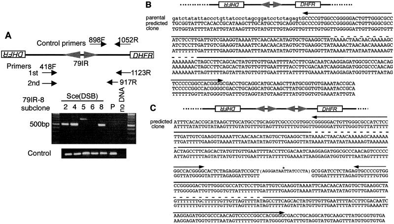
Figure 4.
PCR amplification and sequencing of the bisulfite-modified DNA from the palindromic center. (A) PCR amplification. Nested PCR was used to amplify the junctions of the rearranged DNA. The locations of the primers used, including those for the positive controls, are indicated. DNA from 79IR-8 parental cells (P) and MTX-resistant subclones 2, 4, 5, 6, and 8 were treated with bisulfite and amplified. Subclones 2, 4, and 8 produced an ≈500-bp PCR fragment with the palindromic structure diagrammed. Subclone 5 produced an ≈700-bp fragment. There is no amplification product from subclone 6 or a parental transformant. (B) Sequence of the short IR-mediated palindrome from subclone 2. PCR fragments described above were cloned and sequenced (bottom line, bisulfite modified) and compared with the predicted palindromic sequence (middle line). Bisulfite treatment converts C in the middle lines to T in the bottom line. The parental nonpalindromic sequence in this region is also shown (top line). The sequence upstream of the 79-bp IR is shown in lowercase. The two divergent arrows indicate the locations of the 79-bp IR, and the dashed line indicates the nonpalindromic center. A schematic drawing is shown at the top. (C) Sequence of the end-to-end joining palindrome from subclone 5. The sequence after bisulfite modification (bottom line) was shown along with the predicted sequence (top line). The two palindromic arms are joined head to head at the I-SceI cleaved site (asterisk) with 9 bp and 8 bp of deletions from the two sides (end-to-end joining palindrome), which is shown in the parenthesis. Note that two pairs of the short IR (arrows), including the nonpalindromic center (dashed line), are present in this sequence, compared with only one in the sequence in B.

