Figure 5.
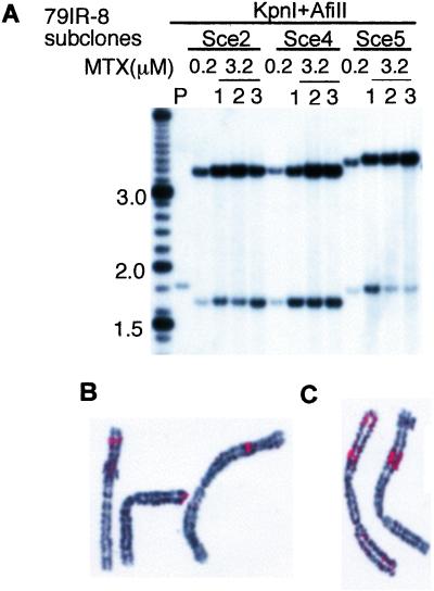
Intrachromosomal amplification of DHFR. (A) Subclones with short IR-mediated palindrome (subclones 2 and 4) and end-to-end joining palindrome (clone 5) isolated in 0.2 μM MTX from transformant 79IR-8 were further selected with 3.2 μM MTX, and resistant subclones were isolated after 10 days of growth. The genomic DNA was digested with KpnI and AfiII and analyzed by Southern blotting. (B) Intrachromosomal palindrome formation of DHFR transgene in subclone 2. Chromosomes shown are from three different cells. FISH was performed by using DNA fragment from pD77IRSce and 5 kb of the flanking genomic sequence as a probe. Each signal (red) probably represents one palindrome of the DHFR transgene. (C) In the highly amplified Sce2–1 subclone, large blocks of signals were seen in different cells on the same chromosome, which, in some cases, showed fused sister chromatids (Left).

