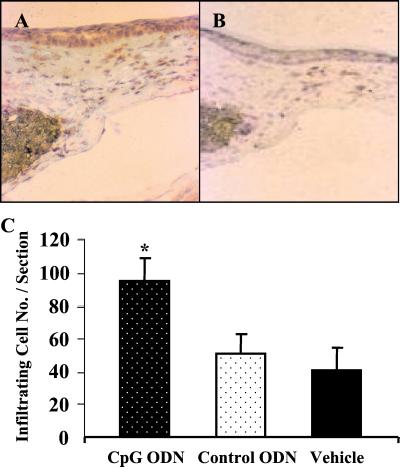
Figure 4.
CpG DNA induces inflammation and VEGF expression in corneal micropockets. Pellets containing 1 μg of CpG (A) or control (B) ODN were implanted into mouse corneas. Frozen sections from these eyes were stained for VEGF-expressing cells (dark-brown stain) 4 days later. Positive cells are present in the ipsilateral site of the pellet-implanted cornea (200×). (C) Cellular infiltration was quantified by enumerating infiltrating cells in the corneal stroma. Each point represents the mean total cellular infiltrate from four central corneal sections from two eyes.

